Teeth names
What every tooth in your mouth is called, what it does, and why understanding it helps you
Every tooth in your mouth has a name, a number and a purpose. Most patients hear these terms at dental appointments and nod along without really knowing what is being referred to. “We are going to look at the upper right first molar.” “The lower left canine has a crack.” “Your second premolars are showing some wear.”
These are not arbitrary labels. Teeth names exist because different teeth have fundamentally different structures, different functions and different clinical vulnerabilities. The treatment appropriate for an incisor is different from the treatment appropriate for a molar. The way each tooth fails, the way each tooth is restored, and the symptoms each tooth produces when something goes wrong all vary significantly depending on which tooth you are talking about.
Understanding what teeth are called also helps you make sense of your own dental records, understand treatment recommendations, and have clearer conversations with your dental team. This guide explains every tooth type clearly, what each one does, why it matters clinically, and how the naming system fits together.
At Lateral Dental Clinic in Sheffield, led by Dr Matthew Stephens GDC No. 263989 and Dr Anupa Stephens GDC No. 264031, we believe informed patients make better decisions about their dental care. Here is the full picture.
How many teeth do adults have, and how are they arranged?
A full adult (permanent) dentition consists of 32 teeth. These are arranged symmetrically: 16 in the upper jaw (maxilla) and 16 in the lower jaw (mandible). Each half of each jaw contains 8 teeth, giving four quadrants of 8 teeth each.
The 32 teeth include four types:
- 8 incisors (4 upper, 4 lower)
- 4 canines (2 upper, 2 lower)
- 8 premolars (4 upper, 4 lower)
- 12 molars (6 upper, 6 lower, including 4 wisdom teeth)
Many adults have fewer than 32 teeth because the third molars (wisdom teeth) have been removed, have failed to erupt, or were never developmentally present. A dentition without wisdom teeth still contains 28 teeth and is entirely functional.
Children have a primary (baby) dentition of 20 teeth: 8 incisors, 4 canines and 8 molars. Primary teeth do not include premolars. These are replaced by the permanent dentition gradually between the ages of approximately 6 and 12.
The numbering systems dentists use
Two numbering systems are used in UK dental practice, and understanding them helps make sense of your dental records.
The FDI (Federation Dentaire Internationale) two-digit system
This is the most widely used system in the UK and internationally. It divides the mouth into four quadrants, numbered 1 to 4 for permanent teeth:
- Quadrant 1: upper right
- Quadrant 2: upper left
- Quadrant 3: lower left
- Quadrant 4: lower right
Within each quadrant, teeth are numbered 1 to 8 from the midline outward. So “11” is the upper right central incisor, “21” is the upper left central incisor, “36” is the lower left first molar, and “47” is the lower right second molar.
For primary teeth, the quadrant numbers are 5, 6, 7 and 8, with tooth positions numbered 1 to 5.
The Universal Numbering System
Used more commonly in the United States but encountered in some UK records and software, this system numbers all 32 permanent teeth from 1 to 32, starting at the upper right third molar and moving clockwise around the mouth.
Both systems are precise and unambiguous, which is why dentists use them: “upper right first molar” can be misunderstood, but “16” is a specific, unambiguous tooth.
Incisors: the cutting teeth
What are incisors?
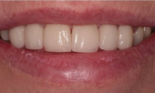
Incisors are the four upper and four lower flat, chisel-shaped teeth at the very front of the mouth. They are the teeth most visible when you smile, most directly involved in speech, and most important for the aesthetics of the smile.
There are two types of incisor in each jaw: central incisors and lateral incisors.
Central incisors are the two teeth at the absolute midline, one on each side. They are the largest and most prominent of the front teeth. The upper central incisors are the teeth that dominate the aesthetic zone and are the primary focus of any smile makeover or cosmetic dental work.
Lateral incisors sit immediately beside the central incisors. They are slightly smaller and narrower. In some patients, the upper lateral incisors are developmentally very small, a condition called peg laterals, which can be addressed with composite bonding or porcelain veneers.
What incisors do
Their primary function is incision: biting off portions of food through a clean, shearing action. They do not grind. Their thin, flat shape and sharp edge are designed specifically for this cutting function.
Incisors also play a critical role in speech, particularly for sounds that involve contact between the tongue and the back of the upper front teeth, and for certain fricative sounds where the lower lip contacts the upper incisors.
Clinically, incisors are:
- The teeth most commonly affected by trauma (chips, fractures, avulsion from falls or impacts)
- The teeth where acid erosion is most visible, particularly on the palatal surfaces of the upper incisors
- The teeth most involved in cosmetic dentistry, including composite bonding, porcelain veneers and teeth whitening
- Among the easier teeth to numb with local anaesthetic due to the thin bone overlying their roots
A cracked or fractured incisor may need composite bonding if minor, a crown if significant, or root canal treatment if the fracture has reached the pulp.
Canines: the cornerstone teeth
What are canines?
Canines are the pointed teeth that sit at the corners of the dental arch, one in each quadrant. They are called canines because of their resemblance to the prominent teeth of carnivorous animals. They are also called cuspids or eye teeth (particularly the upper canines, which sit beneath the eye socket).
There are four canines in a full adult dentition: upper right, upper left, lower right and lower left. In the FDI system they are teeth 13, 23, 33 and 43.
What canines do
Canines have several distinct roles that make them clinically significant beyond their appearance.
Their primary mechanical function is tearing and holding food. The pointed cusp form and the long, robust root make them particularly suited to gripping.
More importantly from a dental function perspective, canines typically provide what is called canine guidance: during lateral (side-to-side) jaw movements, the upper and lower canines on the working side come into contact and cause the back teeth on that side to separate slightly. This separation protects the molars and premolars from the lateral forces of chewing, which they are not designed to absorb routinely.
When canine guidance is absent, either because the canines are worn, missing or mal-positioned, the back teeth take lateral forces they should not receive, leading to accelerated wear, fractures and muscle strain.
Canines also have the longest roots of any tooth in the mouth, making them particularly stable and resistant to loss from gum disease. They are often the last teeth to be lost even in patients with advanced periodontal disease.
Clinically, canines are:
- The teeth most important for guiding the bite during lateral movements
- The teeth most commonly involved in guidance-related wear patterns
- Important anchors for partial dentures
- Resistant to extraction compared to other teeth due to their root length and bony support
Premolars: the transitional grinders
What are premolars?
Premolars, also called bicuspids, sit between the canines and the molars. There are two premolars in each quadrant: first premolar and second premolar. This gives eight premolars in total in a full adult dentition.
In the FDI system, the first premolars are teeth 14, 24, 34 and 44. The second premolars are 15, 25, 35 and 45.
Premolars do not exist in the primary (baby) dentition: children have molars where the adult premolars will eventually erupt.
What premolars do
As their intermediate position suggests, premolars combine the functions of the teeth on either side. They assist the canines with tearing and the molars with grinding. Their two-cusped form (bicuspid refers to the two cusps that most premolars have) provides a surface capable of both shearing and crushing food.
The upper first premolars are the teeth most commonly extracted for orthodontic purposes, particularly when crowding requires space to be created for alignment. Their removal, when planned orthodontically, does not significantly affect biting function because the adjacent teeth adapt to the change.
Clinically, premolars are:
- Among the most commonly affected teeth for a type of wear called abfraction, where stress-related cervical lesions develop at the gum margin
- Prone to crack lines developing within large restorations, particularly when significant tooth structure has been removed by previous fillings
- The teeth most commonly receiving dental crowns following fracture, particularly when the remaining tooth structure is insufficient for a filling alone
- Sometimes the teeth where additional roots are present, which can make root canal treatment more complex than anticipated
Molars: the heavy grinding teeth
What are molars?
Molars are the large, multi-cusped teeth at the back of the mouth. There are three molars in each quadrant of a full adult dentition: first, second and third. The third molar is the wisdom tooth. This gives 12 molars in total, including 4 wisdom teeth.
In the FDI system, the first molars are 16, 26, 36 and 46. The second molars are 17, 27, 37 and 47. The third molars (wisdom teeth) are 18, 28, 38 and 48.
What molars do
Molars are the workhorses of the dentition. Their broad, flat, multi-cusped occlusal (biting) surface is designed for grinding food into small particles before swallowing. They absorb the majority of the biting force generated during chewing.
The lower first molars are typically the first permanent teeth to erupt (around age 6) and are therefore the longest-standing teeth in the adult dentition by the time problems arise. This, combined with their position and the heavy forces they absorb, makes them among the teeth most frequently affected by decay and fracture in adult patients.
What are molars that cause the most clinical problems?
The first molars, particularly the lower first molars. Because they erupt at age 6, they have the longest exposure time to the oral environment, the most cycles of biting force, and in many patients they carry the largest restorations from treatments carried out years earlier. A lower first molar with a large amalgam filling placed in childhood may be significantly cracked, partially fractured and at risk of losing a cusp by the time the patient is in their forties.
The third molars, the wisdom teeth, are discussed separately below.
Clinically, molars are:
- The teeth most frequently requiring dental crowns due to fracture from heavy biting forces combined with large existing restorations
- The teeth with the most complex root anatomy, often having three or four root canals, which makes root canal treatment more involved
- The teeth most affected by cuspal fractures when existing fillings have removed the natural internal buttressing of the tooth walls
- Among the most heavily loaded teeth in bruxism patients, making them a priority for protective coverage with a night guard or crown
Wisdom teeth: the third molars
Wisdom teeth are the four third molars that sit at the very back of each quadrant. They are named for their late arrival: they typically erupt between the ages of 17 and 25, by which time most other decisions about life have already been made.
In many people, wisdom teeth cause problems because there is insufficient space in the jaw to accommodate them. They can:
- Become impacted (unable to erupt into a normal position)
- Partially erupt, leaving a gum flap over the crown that traps bacteria and leads to infection (pericoronitis)
- Erupt at an angle that presses against the adjacent second molar
- Develop decay in a position so inaccessible that restoration is impractical
Not every wisdom tooth requires removal. Wisdom teeth that have erupted fully, are in a good position and can be cleaned effectively contribute to chewing function and do not need to be extracted. Those causing recurrent infection, adjacent tooth damage or significant bone loss are usually best removed.
Primary teeth: the first set
Teeth names in the primary dentition follow the same pattern but with a simplified version. Primary teeth are labelled A through T in some systems, or using the FDI quadrant numbers 5 through 8.
The 20 primary teeth are:
- 8 incisors (central and lateral, same as the permanent set)
- 4 canines (same positions as the adult canines)
- 8 primary molars (in the positions where premolars and first molars will eventually sit)
Primary teeth do not include premolars. The spaces held by the primary molars are eventually taken by the permanent premolars and first molars.
Primary teeth matter more than many parents appreciate. They hold space for the permanent teeth beneath them, support speech development and nutrition during childhood, and when lost prematurely to decay or trauma, the adjacent teeth can drift and reduce the space available for the permanent teeth to erupt. This is why dentists treat primary teeth rather than dismissing concerns with “they are going to fall out anyway.”
Why teeth names matter for your dental health
Understanding what teeth are called helps you take a more active role in your dental care. When a dentist says “the upper right first molar has a hairline crack running toward the pulp and may need a crown and root canal treatment,” the patient who understands what that means can ask better questions, make a more informed decision, and understand what the treatment involves.
It also helps you identify and describe symptoms accurately. “I have a sharp pain in the tooth at the back on the lower right when I bite” is more helpful than “one of my back teeth hurts.”
How to book a check-up at Lateral Dental Clinic, Sheffield
A dental check-up at Lateral Dental Clinic includes a full assessment of every tooth, named and numbered, with X-rays where appropriate and a clear explanation of everything found. For patients who are anxious about dental appointments, conscious sedation is available, allowing treatment to proceed in a state of deep relaxation. You can book an appointment directly online at any time.
In conclusion
Teeth names are not jargon: they are a precise language for talking about specific structures that have specific functions and specific vulnerabilities. Incisors cut. Canines tear and guide the bite. Premolars bridge the two zones. Molars grind. Each type has its own common problems, its own treatment pathways, and its own clinical significance.
Knowing what teeth are called, understanding what molars are, and understanding what canines are gives you the vocabulary to engage fully with your dental care rather than nodding along. And it makes the check-up conversation considerably more useful for both the patient and the clinical team.
Explore our full range of general dentistry and cosmetic services at Lateral Dental Clinic in Sheffield, or book your next check-up online.
Disclaimer
The information in this article is intended for general educational guidance only and does not constitute personalised dental advice. For concerns about specific teeth, please book an appointment with a qualified dental professional for a proper clinical assessment.
Lateral Dental Clinic is a private dental practice in Sheffield, led by Dr Matthew Stephens GDC No. 263989 and Dr Anupa Stephens GDC No. 264031. We offer dental check-ups, root canal treatment, dental crowns, dental sedation, general dentistry, Invisalign, composite bonding, porcelain veneers, teeth whitening and smile makeovers. Book your appointment online.
Frequently asked questions
The four types of teeth in the adult dentition are incisors, canines, premolars and molars. Incisors are the flat front teeth used for cutting. Canines are the pointed corner teeth used for tearing and for guiding the bite. Premolars sit between the canines and molars and combine cutting and grinding functions. Molars are the large back teeth that do the majority of the chewing and grinding work. A full adult dentition has 32 teeth across these four types, including 4 wisdom teeth (third molars).
Canines are the four pointed teeth, one in each corner of the mouth. They are the longest-rooted teeth in the mouth and are clinically significant primarily because of the role they play in guiding the bite: during lateral jaw movements, the upper and lower canines on the same side contact each other and protect the back teeth from lateral forces. When canines are worn down or missing, the molars and premolars absorb forces they are not designed for, leading to accelerated wear and fracture. A thorough dental check-up assesses canine guidance as part of the overall bite assessment.
Molars are the large back teeth responsible for the majority of chewing force. They have broad, multi-cusped surfaces, multiple roots and complex root canal anatomy. Because they absorb such significant force over decades, and because they typically carry large fillings that weaken the remaining tooth structure, they are the teeth most prone to cuspal fracture. A fractured cusp on a molar often requires a dental crown. Where the fracture or previous decay has reached the pulp, root canal treatment is needed before the crown is placed.
Numbers are precise and unambiguous. Saying “the upper right first molar” is accurate but slightly open to interpretation depending on whether the speaker is facing the patient. The FDI two-digit notation, where “16” always refers to the upper right first molar regardless of perspective, removes all ambiguity. It also allows dental records, referral letters and treatment plans to be understood identically by any clinician anywhere in the world. Patients hearing these numbers during appointments can ask what specific tooth is being referred to, and the dental team at Lateral Dental Clinic are always happy to explain.
The same types exist, with the exception that primary teeth do not include premolars. Children have central and lateral incisors, canines, and primary molars in the positions where permanent premolars and first molars will eventually sit. The naming convention is similar but the specific designations differ. Primary teeth are often referred to by letter (A to T in one system) or by their FDI quadrant numbers 5 to 8. Understanding that primary teeth hold space for permanent teeth, support speech and nutrition, and matter clinically in their own right is one of the reasons we recommend dental check-ups for children from a young age. Book online to arrange an appointment for the whole family.